

The recumbent ewe around lambing time can be frustrating for both the farmer and veterinary surgeon, as they can take up a lot of valuable time at a very busy time of year. The most common causes of recumbency in ewes around lambing time are related to nutrition: either as a result of the high metabolic demands of the ewe (for example hypocalcaemia, pregnancy toxaemia and hypomagnesaemia) or feedstuffs (for example listeriosis and rumen acidosis). Careful clinical examination and history is required to determine the most likely cause, although clinical biochemistry can provide cost-effective laboratory confirmation of the diagnosis.
Hypocalcaemia
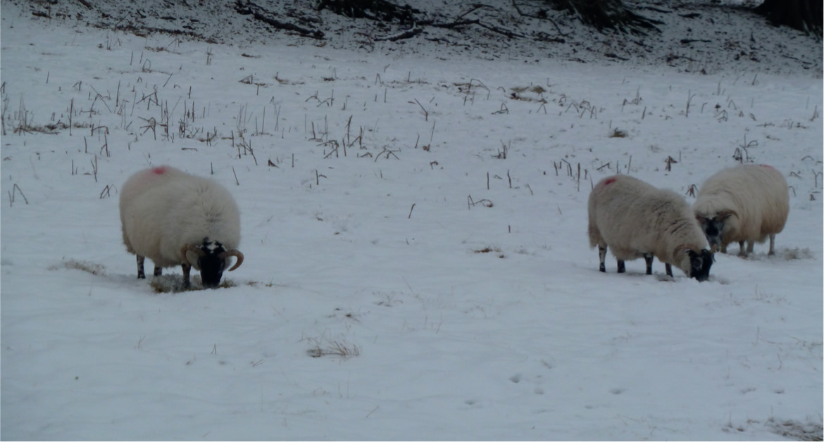
Hypocalcaemia is commonly seen in ewes in late pregnancy (last 6 weeks of gestation), and may also occur sporadically in early lactation. The pathophysiology is similar to that of milk fever in cows, in that it occurs when the calcium requirements for pregnancy and lactation are not met by dietary calcium absorption and mobilisation of skeletal calcium reserves, and there is a failure of blood calcium hormonal regulation mechanisms. It is more common in older sheep (3-crop or older), and ewes in any body condition may be affected. In sheep, it is frequently associated with stressful husbandry events (e.g. housing, movement in-bye, dog worrying, delay/change in feed: Figure 1) and may thus occur as ‘outbreaks’ with multiple ewes affected.
The clinical signs of hypocalcaemia are due to muscular paralysis. Ewes become separated from the flock, showing incoordination, weakness and (rarely) hyperaesthesia. The disease then progresses to give signs of recumbency, depression, bloat and constipation (Cockcroft and Whiteley, 1999). Ewes have a rapid respiratory rate (‘panting’) with passive reflux of ruminal contents down the nose, which can be mistaken by farmers for respiratory disease.
Diagnosis is made on the basis of clinical signs, history of recent stressful husbandry procedures and response to treatment. Serum calcium concentrations below 1.0 mmol/litre support the diagnosis. As affected ewes are frequently inappetant for a period of time during the initial phase of hypocalcaemia, β-OH butyrate (BOHB) levels are commonly elevated and ewes may go on to develop pregnancy toxaemia.
Treatment for a 70 kg ewe is by the administration of 20–40 mls of warm 40% calcium borogluconate by slow intravenous injection, and 50–100 ml subcutaneously (Scott, 1995). As with milk fever in a cow, the ewe should respond within minutes with improvement in demeanour, eructation, defecation, urination and attempts to rise. Although affected cases are also hypophospataemic (serum inorganic phosphate below 0.9 mmol/litre), they will respond to calcium borogluconate alone and thus the clinical relevance of this low phosphate is unclear.
Control and preventative measures for hypocalcaemia in sheep include the avoidance of stressful husbandry conditions such as provision of adequate food (particularly during harsh weather conditions: Figure 1), timing of vaccinations before lambing, avoidance of unnecessary transportation in late pregnancy etc. However such stressors are oft en unavoidable, and so increased farmer vigilance for recumbent ewes and prompt treatment is advisable aft er such husbandry issues arise.
Dietary control of hypocalcaemia in sheep in late pregnancy remains the subject of much debate! Based on the traditional ‘calcium restriction’ method of nutritional manipulation in late pregnancy for the control of milk fever in dairy cows, it was advocated that prelambing diets should contain adequate levels of calcium, but not excessive quantities that will suppress the homeostatic calcium mechanisms (Brozos et al, 2011; Povey et al, 2016). This approach would recommended intakes of 5 g of calcium per day for ewes in late pregnancy. While manipulation of acid–base balance (also termed dietary cation–anion balance: DCAB) is used widely for milk fever control in dairy cows, it has only ever been performed experimentally in sheep, and this approach has never been shown to prevent hypocalcaemia in sheep.
However there is a counter-argument that the pathogenesis of hypocalcaemia in sheep is different to that in dairy cattle, as the disease is more chronic during late pregnancy (compared with the acute changes in calcium requirements that occur at calving in dairy cattle), and is complicated by the provision of poor quality pasture in the late winter period. Anecdotal evidence is that calcium supplementation in late pregnancy may be beneficial in certain situations, and so some feed advisors recommend supplementing with higher levels of calcium in late pregnancy via concentrate feed.
There are also anecdotal reports of breed effects on hypocalcaemia, with hill breeds such as Swaledales and Blackfaces reported to have higher prevalence. Whether this is a true breed susceptibility or affected by the management of hill breeds on poorer quality pastures remains to be determined. The relationship between calcium, phosphorous and magnesium is also likely to be important for calcium homeostasis, with excess levels of phosphorous (and so an excessive dietary calcium:phosphorous ratio less than 1:1) considered a risk factor (Brozos et al, 2011). Oxalate can bind calcium in the diet making it unavailable, and so it is not recommended to feed feedstuffs such as sugar beet tops that contain high levels of oxalate to ewes in late pregnancy. The role of vitamin D is also of interest, as it plays an important role in absorption of calcium from the gut and mobilisation from bone. Low vitamin D status has been linked with the development of bone defects in young lambs (Mearns et al, 2008), and it could be that poor vitamin D status in late pregnancy is associated with an increased risk of hypocalcaemia.
Hypomagnesaemia
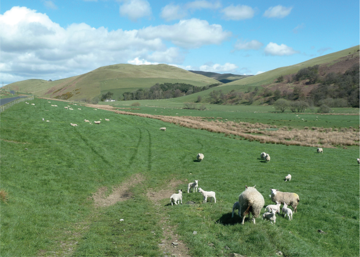
Hypomagnesaemia in sheep nearly always occurs after lambing (usually the first 6 weeks after lambing), in ewes rearing twins at peak lactation as milk production represents the largest drain on magnesium (Figure 2). The usual history is that the ewes are grazing on lush pasture, which have received high applications of artificial fertilizers (high nitrogen and potassium) which reduce magnesium absorption from the gut. There have also been reports of hypomagnesaemia developing secondary to pasture sodium deficiency, as sodium is required for magnesium absorption (Sargison et al, 2004). Unlike the situation with calcium, there are no hormonal control mechanisms to regulate blood magnesium levels, and so blood levels are dependent on dietary intake and absorption from the gastrointestinal tract.
Clinical signs include hyperaesthesia and trembling, incoordination, collapse and tetanic convulsions. The condition is usually sudden in onset, and progresses rapidly. Cases frequently present as sudden death in adult lactating ewes.
While diagnosis is often based on clinical signs, serum magnesium concentrations below 0.6 mmol/litre will help confirm the diagnosis, and distinguish from other cases of recumbency. Investigation of hypomagnesaemia as a cause of sudden death can be confirmed by fresh cerebrospinal fluid (CSF) and urine magnesium concentrations below 0.6 mmol/litre, or fresh aqueous humour magnesium concentrations below 0.5 mmol/litre (within 24 hours of death)(McCoy et al, 2001).
Treatment of hypomagnesaemia for a 70 kg ewe involves the administration of 20 mls of warm 20% calcium borogluconate and magnesium solution intravenously, and 50 mls of warm 25% magnesium sulphate subcutaneously. Mortality rates can be high unless treatment is very rapid.
Lactating ewes that are out at grass require supplementation with magnesium to ensure target intakes of 7 g of magnesium per ewe per day. The most reliable way to perform this with ewes in lactation at grass is via concentrates, which usually contain 14 g of magnesium oxide (calcined magnestite).
There are magnesium boluses available for sheep (e.g. Rumbul Sheep™, Agrimin), although they are supplementary rather than supplying all of the ewes' magnesium requirements. Providing supplementary forage such as big bale silage will increase gut transit time and so increase magnesium absorption, but intakes are likely to be variable when good quality grazing is available. Reducing stress (such as provision of shelter) will also help reduce the risk of clinical disease developing.
Pregnancy toxaemia (twin-lamb disease)
Pregnancy toxaemia is a commonly encountered metabolic disease in lowground flocks in the last month of pregnancy. The rapid growth of the fetus in late pregnancy results in a marked increase in glucose requirements of the gravid uterus and so the heavily pregnant ewe thus has to meet the energy demands of the growing fetus, as well as her own maintenance demands. This problem is exacerbated by the decreased dry matter intake in late pregnancy due to the increased volume of the pregnant uterus. The hormonal balance in late gestation (decreased insulin; increased growth hormone, prolactin and progesterone) gives priority to the glucose needs of the fetus over the needs for maintenance, leading to fat mobilisation and hyoglycaemia.
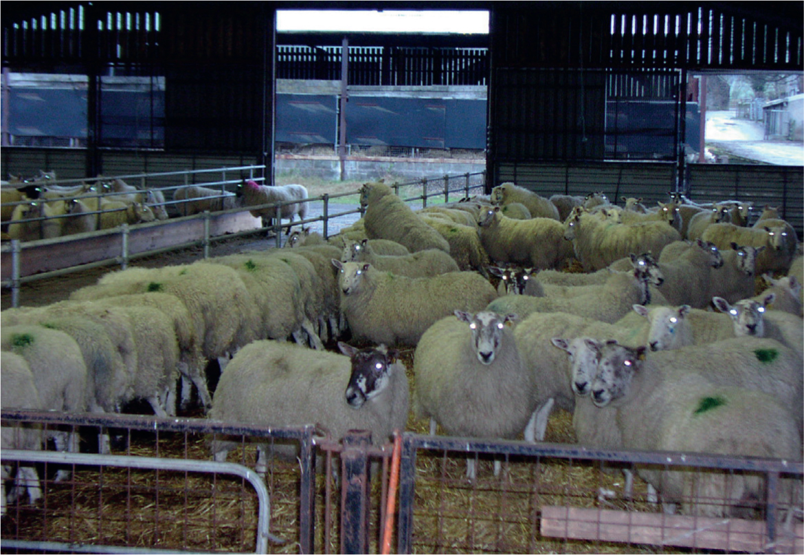
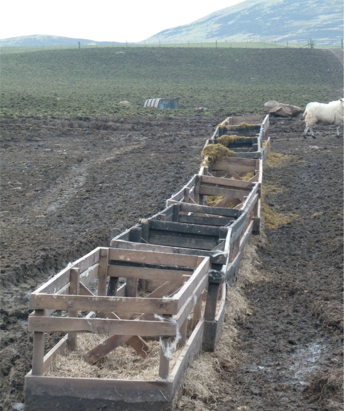
Predisposing factors for the development of pregnancy toxaemia include: ewes carrying two or more lambs; a prolonged period of energy shortage (such poor forage quality, inadequate concentrates, poor feed intakes, high fetal demand: Figure 3); ewes in poor body condition score (BCS, below 2) that have little body reserves to meet any shortfall in energy demand; overfat ewes (over BCS 4) that are prone to excessive fat mobilisation and the development of fatty liver; older ewes (due to other problems such as broken mouth); stressful events such as adverse weather, housing, handling etc. The incidence of other diseases such as vaginal prolapse, hypocalcaemia, lameness etc. may also precipitate cases of pregnancy toxaemia due to periods of inappetance in late pregnancy.
Initial clinical signs include isolation from the remainder of the flock, and refusal to feed. The ewes are easy to catch, and become increasingly dull and depressed. Neurological signs then progress because of the development of hypoglycaemic encephalopathy. The ewes are apparently blind, with lack of menace response but normal pupillary light response. Fine tremors of the head, neck and ears with hyperaesthesia develop, as well as abnormal behaviours such as head pressing, star-gazing, teeth grinding and incoordination. This then progresses over a period of days to weakness, profound depression and recumbency. It is characterised by a high mortality rate (over 90% of affected ewes will die if untreated). Affected ewes will frequently abort the fetuses, which will often result in a marked improvement of the ewe's condition.
Diagnosis is frequently made based on clinical signs and history. Confirmation of diagnosis is by serum BOHB levels over 3.0 mmol/litre. Approximately 25% of affected ewes will also be hypocalcaemic, and ewes with advanced liver damage will have elevated liver enzymes (such as aspartate aminotransferase (AST) and glutamate dehydrogenase (GLDH)). Post-mortem examination will reveal fatty infiltration of the liver, and the presence of two or more fetuses in the uterus. Post-mortem diagnosis can be confirmed by aqueous humour BOHB levels above 2.5 mmol/litre.
Response to treatment tends to be poor, with approximately 70–80% mortality rates usually quoted. Treatment must be instigated early to have any chance of success. Initial treatment with 100 ml of 40% dextrose intravenously will temporarily restore blood glucose levels, and 50 ml propylene glycol orally four times daily will also supply proprionate precursors to help with energy metabolism. Affected ewes should be removed from the group, offered concentrate feeds and water, good bedding and shelter. Multivitamins and small doses of steroids (5 mg dexamethasone, which should not be sufficient to induce parturition) are also given by some veterinary surgeons in practice to improve liver metabolism and gluconeogenesis.
An alternative treatment strategy is to remove the fetal energy demands by inducing parturition. If the ewe is more than 136 days pregnant, induction of parturition can be performed using 16 mg dexamethasone, and will take approximately 48 hours. Alternatively an elective Caesarean operation may be considered. In advanced cases, euthanasia of the ewe should be considered.
Control and preventative measures are dependent on the correct nutrition of the ewe in late pregnancy (Macrae, 2017).
Listeriosis
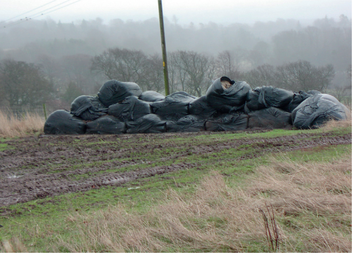
The nervous form of listeriosis is commonly encountered in ewes in late pregnancy being fed poor quality silage. It is primarily a sporadic condition affecting small numbers of individual ewes, although outbreaks can occur within a flock. Risk factors for high levels of Listeria monocytogenes multiplication in silage include poor fermentation (silage pH >5), soil contamination (which usually occurs when the silage was made in wet conditions) and aerobic spoilage (due to holes in silage wrap or clamp covers). Feeding big bale silage tends to have the highest risk (Figure 4), although clamp silage can be a risk especially if feeding practices are poor allowing secondary fermentation and spoilage. Listeria spp. outbreaks have also been reported that are not linked to silage feeding, but have usually involved soil contamination and/or feeding poorly preserved feedstuffs.
Affected sheep are often younger animals (such as gimmers) due to the changing of their teeth, which allows L. monocytogenes to enter the trigeminal nerve in the mouth. Initial clinical signs include isolation from the remainder of flock, and refusal to feed. The ewes are easy to catch, and become increasingly dull and depressed. They often propel themselves into corners or under gates. Classically affected sheep will display unilateral facial nerve paralysis, including drooping of the ear and eyelid, deviation of the muzzle to the unaffected side, flaccid lip, impaction of food in the cheek and drooling of saliva. Other cranial nerves may be affected, and circling behaviour may occur if the vestibular-cocchlear nucleus is affected. Deterioration occurs rapidly over a couple of days without treatment.
Diagnosis in the live animal is usually made on the basis of clinical signs, following a detailed neurological assessment. Difficulty in confirming the clinical diagnosis occurs when signs of cranial nerve deficits are slight or bilateral. CSF sampling may assist in achieving a diagnosis (Scott, 1993).
Treatment involves high doses of penicillin G (300 000 IU/kg intravenously on day 1, following by 44 000 IU/kg intramuscularly daily for 5 days: Scott 2007). However treatment success rates are often disappointing, with 70% mortality rates often quoted (Scott, 2007). Affected ewes should be removed from the group, offered concentrate feeds and water, good bedding and shelter. They may require assistance with feeding and nursing due to difficulties in eating and swallowing.
Prevention and control involves feeding good quality silage that has been produced and stored under conditions that do not allow the multiplication of L. monocytogenes. Spoiled silage should be discarded, and fed to other lower-risk groups such as growing cattle. Fresh silage should be fed each day, with refusals cleared away to prevent the risk of secondary fermentation, and contamination of silage with soil or manure should be minimised (Figure 5).
Other potential causes of recumbency
There are a number of other potential causes of recumbency in ewes in late pregnancy. These would tend to be less common over-all, but may be significant in individual flocks and so need to be considered:
- Ruminal acidosis — this occurs as a result of carbohydrate overfeeding, which can be encountered in lowground flocks during late pregnancy when high levels of concentrate may be fed to twins and triplets. Disruptions in feed supply and insufficient trough space may compound problems (Figure 3), when individual ewes may eat more than their requirements. Clinical examination often reveals the presence of diarrhoea with undigested grains in the faeces, as well as rumen bloat.
- Copper poisoning — this may occur in susceptible breeds such as Beltex, Texels or Bluefaced Leicesters, either as a result of prolonged feeding of concentrates (high in background levels of copper) or feeding inappropriate minerals (such as cattle minerals). Clinical examination will reveal the presence of jaundice and haemolytic anaemia.
- Polioencephalomalacia (cerebrocortical necrosis) — more commonly seen in weaned lambs in the autumn, it can also occur in ewes following sudden changes in diet during late pregnancy (such as housing or introduction of concentrate feeding). Affected animals are blind, and characteristically often display opisthotonus or ‘star gazing’. Without appropriate treatment with vitamin B1 (thiamine), the condition deteriorates to recumbency, seizure activity and death within a couple of days.
- Other neurological disease (e.g. vestibular lesions, brain abscess, coenurosis). These may present with a variety of clinical signs including recumbency, but will usually only affect individual animals.
- Severe emaciation as a result of prolonged malnutrition or disease (for example chronic liver fluke), which would be seen with ewes in BCS 1.5 or below.
Approach to recumbent ewe and suggested ‘first line’ treatment
- Perform a full clinical examination and history. This will give you a guide as to possible diagnosis: listeriosis cases would usually have a history of silage feeding, hypocalcaemia and pregnancy toxaemia predominantly occur prior to lambing, hypomagnesaemia predominantly occurs after lambing. Not all cases obey the textbooks!
- Perform a through and detailed neurological examination, including assessment of cranial nerve reflexes. Be aware that some listeriosis cases may be bilateral, or affect cranial nerves other than facial nerve paralysis.
- Take a pre-treatment blood sample (either serum red top or plasma green top vacutainor) for the assessment of BOHB, calcium and magnesium levels.
- Depending on previous farmer treatment, give 50–100 ml of 20% calcium/magnesium/dextrose solution subcutaneously. Some practitioners also administer 20–40 ml of 20% calcium/magnesium/dextrose solution intravenously, although this route should be used with caution.
- Give additional treatments as directed by the most likely diagnosis on your clinical examination. Note that most diseases in late pregnancy will result in secondary pregnancy toxaemia if the ewe has been inappetant for longer than 12–24 hours, and so oral propylene glycol is likely to be indicated.
- If the ewe fails to respond to treatment, then analysis of the pre-treatment blood sample for BOHB, calcium and magnesium will help with diagnosis.
Assessment of nutritional status in late pregnancy
Prevention is undoubtedly better than cure for most of the main causes of recumbency in ewes around lambing time, especially given the poor treatment success rates of conditions such as pregnancy toxaemia, hypomagnesaemia and listeriosis. Correct nutrition of the ewe in late pregnancy is critical for reducing the risk of such conditions, and further details of late pregnancy nutrition and monitoring can be found in Macrae (2017).
Conclusions
Recumbency in the ewe around lambing time can present a diagnostic challenge to both the farmer as well as the veterinary practitioner. However, a careful clinical examination and history, backed up by appropriate targeted biochemical testing, can help achieve a diagnosis. This allows the development of a treatment plan for recumbent ewes, as well as the implementation of appropriate preventative measures.
KEY POINTS
- Detailed clinical and neurological examination is vital in all recumbent ewes.
- Take a pre-treatment blood sample, for subsequent analysis of β-OH butyrate (BOHB), calcium and magnesium if necessary.
- Most recumbent ewes in late gestation are at risk of developing secondary pregnancy toxaemia.
- Preventative control measures involve correct feed and management of ewes in late pregnancy.