
Taylor & Francis announce the publication of their new textbook Illustrated Textbook of Clinical Diagnosis in Farm Animals which results from a request by undergraduate veterinary students for a ‘5 minute’ reference text that ranks common ruminant diseases showing particular clinical signs or syndromes, such as weakness, tachypnoea/frequent coughing, weight loss, and abdominal distension. The book is designed to complement standard veterinary textbooks and features a comprehensive library of over 1000 video recordings to show how common diseases and disorders typically present on UK farms. Chapters describe clinical examination of each body system with emphasis on key diagnostic tests such as ultrasonography, lumbar cerebrospinal fluid collection and digital radiography and how these can be applied in a cost-effective manner.
For example, published research clearly demonstrates that a sheep's chest can be ultrasound scanned within 30 seconds providing much more accurate information than 5 minutes’ chest auscultation and percussion. The author has used these techniques on farm for the past 30 years yielding many refereed scientific publications and the important findings are reproduced here in the large number of diagnostic video recordings.
The common diseases that present with a particular clinical sign, such as tenesmus, lameness and pruritus are ranked in order of presentation to the author with key clinical signs highlighted using a seven point ‘traffic light’ scale. Images for each disease, diagnostic tests such as sonograms and radiographs, and pathology findings are included. Key observations and clinical examination findings are highlighted in the image legends alongside likely key history events/predisposing factors. Video recordings are used extensively throughout the book to illustrate clinical, ultrasonography and pathology findings. The important laboratory tests are listed in an accompanying table.
Taylor & Francis will provide free access to one book chapter and accompanying video recordings for the next 3 months allowing veterinary undergraduate students to peruse this exciting new approach, especially the extensive repository of unique video recordings. Alongside this offer, the author, Phil Scott, has written a short clinical quiz based on cases featured in this chapter; the winning entry from each UK veterinary school will receive a free copy of the book. The book chapter, links to videos, and answer sheet can be accessed at https://www.routledge.com/9780367612702.
TAYLOR & FRANCIS QUIZ
1.
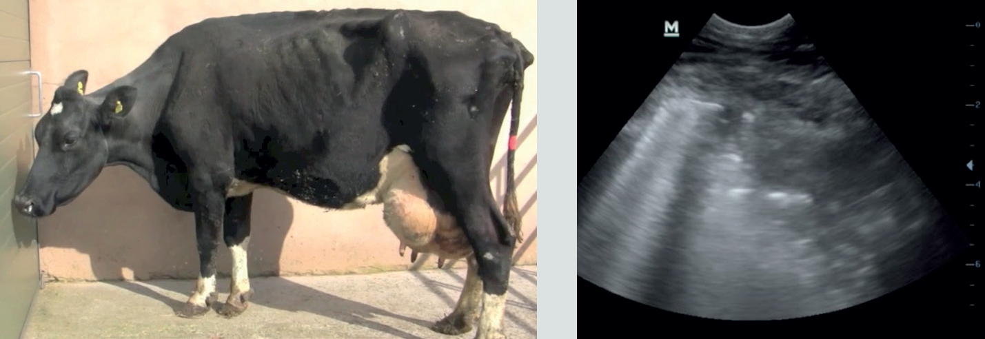
Holstein cow
This recently-calved 6-year-old Holstein cow presents with a much reduced milk yield (11 litres/day), poor appetite, rectal temperature 39.0°C, bilateral purulent nasal discharge, halitosis, an increased respiratory rate and frequent productive coughing. Ultrasound examination of the chest using a 6.5 MHz micro-array probe reveals the far right image.
- Describe the sonographic findings.
- What is the likely cause of the halitosis and frequent productive coughing?
- What it the most likely diagnosis?
- What is the likely best course of action?
2.
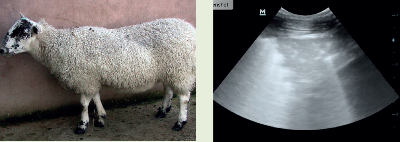
Greyface ewe
During October, the sonogram (right) was obtained by scanning the left ventral chest of a 4-year-old Greyface ewe at the fifth intercostal space using a 6.5 MHz micro-array probe attached to a B-mode ultrasound machine. Dorsal to the left; cm gradations on right margin, probe at top of image.
- Describe the lesion.
- What it the most likely diagnosis?
- What action is necessary?
- What other test could be performed and what is the accuracy of this test?
3.
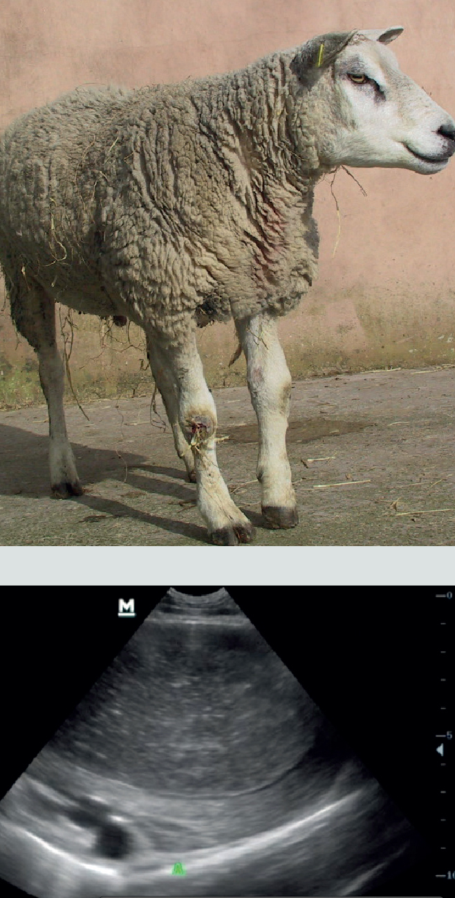
Texel ram
During May, this 4-year-old Texel ram was presented for occasional coughing and an increased respiratory rate observed during gathering. The sonogram (below) was obtained by scanning the right ventral chest at the fifth intercostal space using a 6.5 MHz micro-array probe attached to a B-mode ultrasound machine. Dorsal to the left; cm gradations on right margin, probe at top of image.
- Describe the lesion.
- What it the most likely diagnosis?
- What sounds would be auscultated over this region?
- What is the prognosis for this sheep?
4.
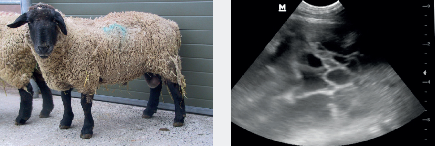
Suffolk ram
The sonogram (below right) was obtained by scanning the ventral chest of a 3-year-old Suffolk ram with a week-long history of poor appetite and an increased respiratory rate with frequent coughing. 6.5 MHz micro-array probe attached to a B-mode ultrasound machine positioned over the fifth intercostal space on right hand side. Dorsal to the left; cm gradations on right margin, probe at top of image.
- Describe the lesion.
- What it the most likely diagnosis?
- What sounds would be auscultated over this lesion?
- What is the prognosis?