References

Agerholm JS. Coxiella burnetii associated reproductive disorders in domestic animals-a critical review. Acta Vet Scand. 2013; 55:(1) https://doi.org/10.1186/1751-0147-55-13

Ahmadinezhad M, Mounesan L, Doosti-Irani A, Behzadi MY. The prevalence of Q fever in the Eastern Mediterranean region: a systematic review and meta-analysis. Epidemiol Health. 2022; 44 https://doi.org/10.4178/epih.e2022097

Amit S, Shinar S, Halutz O, Atiya-Nasagi Y, Giladi M. Suspected person-to-person transmission of Q fever among hospitalized pregnant women. Clin Infect Dis. 2014; 58:(11)e146-e147 https://doi.org/10.1093/cid/ciu151

Arricau-Bouvery N, Rodolakis A. Is Q Fever an emerging or re-emerging zoonosis?. Vet Res. 2005; 36:(3)327-349 https://doi.org/10.1051/vetres:2005010

Astobiza I, Barandika JF, Hurtado A, Juste RA, García-Pérez AL. Kinetics of Coxiella burnetii excretion in a commercial dairy sheep flock after treatment with oxytetracycline. Vet J. 2010; 184:(2)172-175 https://doi.org/10.1016/j.tvjl.2009.01.017

Baud D, Peter O, Langel C, Regan L, Greub G. Seroprevalence of Coxiella burnetii and Brucella abortus among pregnant women. Clin Microbiol Infect. 2009; 15:(5)499-501 https://doi.org/10.1111/j.1469-0691.2009.02779.x

Boni M, Davoust B, Tissot-Dupont H, Raoult D. Survey of seroprevalence of Q fever in dogs in the southeast of France, French Guyana, Martinique, Senegal and the Ivory Coast. Vet Microbiol. 1998; 64:(1)1-5 https://doi.org/10.1016/s0378-1135(98)00247-8

Boone I, Henning K, Hilbert A Are brucellosis, Q fever and melioidosis potential causes of febrile illness in Madagascar?. Acta Trop. 2017; 172:255-262 https://doi.org/10.1016/j.actatropica.2017.05.013

Celina SS, Cerný J. Coxiella burnetii in ticks, livestock, pets and wildlife: A mini-review. Front Vet Sci. 2022; 9 https://doi.org/10.3389/fvets.2022.1068129

Cherry CC, Nichols Heitman K, Bestul NC, Kersh GJ. Acute and chronic Q fever national surveillance – United States, 2008–2017. Zoonoses Public Health. 2022; 69:(2)73-82 https://doi.org/10.1111/zph.12896

Chitanga S, Simulundu E, Simuunza MC First molecular detection and genetic characterization of Coxiella burnetii in Zambian dogs and rodents. Parasit Vectors. 2018; 11:(1) https://doi.org/10.1186/s13071-018-2629-7

Cooper A, Hedlefs R, Ketheesan N, Govan B. Serological evidence of Coxiella burnetii infection in dogs in a regional centre. Aust Vet J. 2011; 89:(10)385-387 https://doi.org/10.1111/j.1751-0813.2011.00819.x

Davies TR, Edwards Y, Morgan A, Caul EO. Prevalence of Q fever in a rural practice. J Public Health (Bangkok). 1997; 19:(3)324-327 https://doi.org/10.1093/oxfordjournals.pubmed.a024638

Davis GE, Cox HR, Parker RR, Dyer RE. A filter-passing infectious agent isolated from ticks. I. Isolation from dermacentor andersonii, reactions with animals, and filtration experiments. Public Health Reports (1896-1970). 1938; 53:(52)2259-2282 https://doi.org/10.2307/4582746

De Biase D, Costagliola A, Del Piero F Coxiella burnetii in Infertile Dairy Cattle With Chronic Endometritis. Vet Pathol. 2018; 55:(4)539-542 https://doi.org/10.1177/0300985818760376

De Cremoux R, Rousset E, Touratier A Assessment of vaccination by a phase I Coxiella burnetii-inactivated vaccine in goat herds in clinical Q fever situation: 1. FEMS Immunol Med Microbiol. 2012; 64:(1)104-106 https://doi.org/10.1111/j.1574-695X.2011.00892.x

Department for Environment, Food and Rural Affairs. New disease reporting requirements from 21st April 2021. http://apha.defra.gov.uk/documents/news/New-disease-reporting-requirements.pdf (accessed 17 August 2023)

Derrick EH. Q “ fever, a new fever entity : clinical features, diagnosis and laboratory investigation. Med J Aust. 1937; 2:(8)281-299 https://doi.org/10.5694/j.1326-5377.1937.tb43743.x

Eldin C, Mélenotte C, Mediannikov O From Q fever to coxiella burnetii infection: A paradigm change. Clin Microbiol Rev. 2017; 30:(1)115-190 https://doi.org/10.1128/CMR.00045-16

Flint J, Dalton CB, Merritt TD Q fever and contact with kangaroos in New South Wales. Commun Dis Intell Q Rep. 2016; 40:(2)E202-E203

Gale P, Kelly L, Mearns R, Duggan J, Snary EL. Q fever through consumption of unpasteurised milk and milk products - a risk profile and exposure assessment. J Appl Microbiol. 2015; 118:(5)1083-1095 https://doi.org/10.1111/jam.12778

Garcia-Ispierto I, López-Helguera I, Tutusaus J Coxiella burnetii shedding during the peripartum period and subsequent fertility in dairy cattle. Reprod Domest Anim. 2013; 48:(3)441-446 https://doi.org/10.1111/rda.12095

Gikas A, Kokkini S, Tsioutis C. Q fever: clinical manifestations and treatment. Expert Rev Anti Infect Ther. 2010; 8:(5)529-539 https://doi.org/10.1586/eri.10.29

Guatteo R, Beaudeau F, Joly A, Seegers H. Coxiella burnetii shedding by dairy cows. Vet Res. 2007; 38:(6)849-860 https://doi.org/10.1051/vetres:2007038

Guatteo R, Seegers H, Joly A, Beaudeau F. Prevention of Coxiella burnetii shedding in infected dairy herds using a phase I C. burnetii inactivated vaccine. Vaccine. 2008; 26:(34)4320-4328 https://doi.org/10.1016/j.vaccine.2008.06.023

Halsby KD, Kirkbride H, Walsh AL The Epidemiology of Q Fever in England and Wales 2000-2015. Vet Sci. 2017; 4:(2) https://doi.org/10.3390/vetsci4020028

Harvey MS, Forbes GB, Marmion BP. An outbreak of Q fever in East Kent. Lancet. 1951; 258:(6695)1152-1157 https://doi.org/10.1016/S0140-6736(51)93157-1

Hawker JI, Ayres JG, Blair I A large outbreak of Q fever in the West Midlands: windborne spread into a metropolitan area?. Commun Dis Public Health. 1998; 1:(3)180-187

Hemsley CM, Essex-Lopresti A, Chisnall T MLVA and com1 genotyping of Coxiella burnetii in farmed ruminants in Great Britain. Vet Microbiol. 2023; 277 https://doi.org/10.1016/j.vetmic.2022.109629

Kampschreur LM, Wegdam-Blans MC, Thijsen SF Acute Q fever related inhospital mortality in the Netherlands. Neth J Med. 2010; 68:(12)408-413

Kersh GJ, Priestley R, Massung RF. Stability of Coxiella burnetii in stored human blood. Transfusion. 2013; 53:(7)1493-1496 https://doi.org/10.1111/j.1537-2995.2012.03912.x

Kopecny L, Bosward KL, Shapiro A, Norris JM. Investigating Coxiella burnetii infection in a breeding cattery at the centre of a Q fever outbreak. J Feline Med Surg. 2013; 15:(12)1037-1045 https://doi.org/10.1177/1098612X13487360

Körner S, Makert GR, Ulbert S, Pfeffer M, Mertens-Scholz K. The prevalence of coxiella burnetii in hard ticks in europe and their role in q fever transmission revisited-a systematic review. Front Vet Sci. 2021; 8 https://doi.org/10.3389/fvets.2021.655715

Kosatsky T. Household outbreak of Q-fever pneumonia related to a parturient cat. Lancet. 1984; 324:(8417-8418)1447-1449 https://doi.org/10.1016/S0140-6736(84)91633-7

Lambton SL, Smith RP, Gillard K, Horigan M, Farren C, Pritchard GC. Serological survey using ELISA to determine the prevalence of Coxiella burnetii infection (Q fever) in sheep and goats in Great Britain. Epidemiol Infect. 2016; 144:(1)19-24 https://doi.org/10.1017/S0950268815000874

Langley JM, Marrie TJ, Covert A, Waag DM, Williams JC. Poker players' pneumonia. An urban outbreak of Q fever following exposure to a parturient cat. N Engl J Med. 1988; 319:(6)354-356 https://doi.org/10.1056/NEJM198808113190607

López-Helguera I, López-Gatius F, Tutusaus J, Garcia-Ispierto I. Reproductive performance of high producing lactating cows in Coxiella-infected herds following vaccination with phase-I Coxiella burnetii vaccine during advanced pregnancy. Vaccine. 2013; 31:(30)3046-3050 https://doi.org/10.1016/j.vaccine.2013.04.067

Ma GC, Norris JM, Mathews KO New insights on the epidemiology of Coxiella burnetii in pet dogs and cats from New South Wales, Australia. Acta Trop. 2020; 205 https://doi.org/10.1016/j.actatropica.2020.105416

Malo JA, Colbran C, Young M An outbreak of Q fever associated with parturient cat exposure at an animal refuge and veterinary clinic in southeast Queensland. Aust N Z J Public Health. 2018; 42:(5)451-455 https://doi.org/10.1111/1753-6405.12784

Marrie TJ. Q fever pneumonia. Infect Dis Clin North Am. 2010; 24:(1)27-41 https://doi.org/10.1016/j.idc.2009.10.004

Marrie TJ, Durant H, Williams JC, Mintz E, Waag DM. Exposure to parturient cats: a risk factor for acquisition of Q fever in Maritime Canada. J Infect Dis. 1988; 158:(1)101-108 https://doi.org/10.1093/infdis/158.1.101

Maurin M, Raoult D. Q fever. Clin Microbiol Rev. 1999; 12:(4)518-553 https://doi.org/10.1128/CMR.12.4.518

McCaughey C, McKenna J, McKenna C Human seroprevalence to Coxiella burnetii (Q fever) in Northern Ireland. Zoonoses Public Health. 2008; 55:(4)189-194 https://doi.org/10.1111/j.1863-2378.2008.01109.x

McCaughey C, Murray LJ, McKenna JP Coxiella burnetii (Q fever) seroprevalence in cattle. Epidemiol Infect. 2010; 138:(1)21-27 https://doi.org/10.1017/S0950268809002854

Mee JF. Investigation of bovine abortion and stillbirth/perinatal mortality - similar diagnostic challenges, different approaches. Ir Vet J. 2020; 73:(1) https://doi.org/10.1186/s13620-020-00172-0

Meredith AL, Cleaveland SC, Denwood MJ, Brown JK, Shaw DJ. Coxiella burnetii (q-fever) seroprevalence in prey and predators in the united kingdom: evaluation of infection in wild rodents, foxes and domestic cats using a modified elisa. Transbound Emerg Dis. 2015; 62:(6)639-649 https://doi.org/10.1111/tbed.12211

Milazzo A, Hall R, Storm PA, Harris RJ, Winslow W, Marmion BP. Sexually transmitted Q fever. Clin Infect Dis. 2001; 33:(3)399-402 https://doi.org/10.1086/321878

Mori M, Roest HJ. Farming, Q fever and public health: agricultural practices and beyond. Arch Public Health. 2018; 76 https://doi.org/10.1186/s13690-017-0248-y

Morroy G, Keijmel SP, Delsing CE Fatigue following acute q-fever: A systematic literature review. PLoS One. 2016a; 11:(5) https://doi.org/10.1371/journal.pone.0155884

Morroy G, Peters JB, van Nieuwenhof M The health status of Q-fever patients after long-term follow-up. BMC Infect Dis. 2011; 11:(1) https://doi.org/10.1186/1471-2334-11-97

Morroy G, Van Der Hoek W, Nanver ZD The health status of a village population, 7 years after a major Q fever outbreak. Epidemiol Infect. 2016b; 144:(6)1153-1162 https://doi.org/10.1017/S0950268815002472

Nusinovici S, Hoch T, Brahim ML, Joly A, Beaudeau F. The effect of wind on coxiella burnetii transmission between cattle herds: A mechanistic approach. Transbound Emerg Dis. 2017; 64:(2)585-592 https://doi.org/10.1111/tbed.12423

Oei W, Kretzschmar MEE, Zaaijer HL, Coutinho R, van der Poel CL, Janssen MP. Estimating the transfusion transmission risk of Q fever. Transfusion. 2014; 54:(7)1705-1711 https://doi.org/10.1111/trf.12539

Patsatzis DG, Wheelhouse N, Tingas EA. Modelling the transmission of coxiella burnetii within a uk dairy herd: investigating the interconnected relationship between the parturition cycle and environment contamination. Vet Sci. 2022; 9:(10) https://doi.org/10.3390/vetsci9100522

Plummer PJ, McClure JT, Menzies P, Morley PS, Van den Brom R, Van Metre DC. Management of C oxiella burnetii infection in livestock populations and the associated zoonotic risk: A consensus statement. J Vet Intern Med. 2018; 32:(5)1481-1494 https://doi.org/10.1111/jvim.15229

Pollock KG, Mellor DJ, Browning LM, Wilson L, Donaghy M. Q fever in migrant workers, Scotland. Emerg Infect Dis. 2007; 13:(12)1963-1964 https://doi.org/10.3201/eid1312.071058

Pommier de Santi V, Briolant S, Mahamat A Q fever epidemic in Cayenne, French Guiana, epidemiologically linked to three-toed sloth. Comp Immunol Microbiol Infect Dis. 2018; 56:34-38 https://doi.org/10.1016/j.cimid.2017.12.004

Pritchard GC, Smith RP, Errington J, Hannon S, Jones RM, Mearns R. Prevalence of Coxiella burnetii in livestock abortion material using PCR. Vet Rec. 2011; 169:(15) https://doi.org/10.1136/vr.d4693

Rabaza A, Fraga M, Corbellini LG, Turner KME, Riet-Correa F, Eisler MC. Molecular prevalence of Coxiella burnetii in bulk-tank milk from bovine dairy herds: Systematic review and meta-analysis. One Health. 2020; 12 https://doi.org/10.1016/j.onehlt.2020.100208

Raoult D, Marrie TJ, Mege JL. Natural history and pathophysiology of Q fever. Lancet Infect Dis. 2005; 5:(4)219-226 https://doi.org/10.1016/S1473-3099(05)70052-9

Raoult D, Stein A. Q fever during pregnancy--a risk for women, fetuses, and obstetricians. N Engl J Med. 1994; 330:(5) https://doi.org/10.1056/NEJM199402033300518

Roest HJ, van Gelderen B, Dinkla A, Frangoulidis D, van Zijderveld F, Rebel J, van Keulen L. Q fever in pregnant goats: pathogenesis and excretion of Coxiella burnetii. PLoS One. 2012; 7:(11) https://doi.org/10.1371/journal.pone.0048949

Taurel AF, Guatteo R, Lehebel A, Joly A, Beaudeau F. Vaccination using phase I vaccine is effective to control Coxiella burnetii shedding in infected dairy cattle herds. Comp Immunol Microbiol Infect Dis. 2014; 37:(1)1-9 https://doi.org/10.1016/j.cimid.2013.10.002

Tilburg JJHC, Roest HJIJ, Buffet S Epidemic genotype of Coxiella burnetii among goats, sheep, and humans in the Netherlands. Emerg Infect Dis. 2012a; 18:(5)887-889 https://doi.org/10.3201/eid1805.111907

Tilburg JJHC, Rossen JWA, van Hannen EJ Genotypic diversity of Coxiella burnetii in the 2007-2010 Q fever outbreak episodes in The Netherlands. J Clin Microbiol. 2012b; 50:(3)1076-1078 https://doi.org/10.1128/JCM.05497-11

van den Brom R, Engelen E, Roest HIJ, Hoek W, Vellema P. Coxiella burnetii infections in sheep or goats: an opinionated review. Vet Microbiol. 2015a; 181:(1-2)119-129 https://doi.org/10.1016/j.vetmic.2015.07.011

van den Brom R, Roest HJ A probably minor role for land-applied goat manure in the transmission of Coxiella burnetii to humans in the 2007-2010 Dutch Q fever outbreak. PLoS One. 2015b; 10:(3) https://doi.org/10.1371/journal.pone.0121355

van der Hoek W, Schneeberger PM, Oomen T Shifting priorities in the aftermath of a Q fever epidemic in 2007 to 2009 in The Netherlands: from acute to chronic infection. Euro Surveill. 2012; 17:(3)

van Leuken JPG, van de Kassteele J, Sauter FJ Improved correlation of human Q fever incidence to modelled C. burnetii concentrations by means of an atmospheric dispersion model. Int J Health Geogr. 2015; 14:(1) https://doi.org/10.1186/s12942-015-0003-y

van Roeden SE, Reukers DFM, van Jaarsveld CHM Chronic Q fever: patient and treatment-related factors influencing long-term quality of life. QJM. 2018; 111:(11)791-797 https://doi.org/10.1093/qjmed/hcy171

van Woerden HC, Mason BW, Nehaul LK Q fever outbreak in industrial setting. Emerg Infect Dis. 2004; 10:(7)1282-1289 https://doi.org/10.3201/eid1007.030536

Velasova M, Damaso A, Prakashbabu BC Herd-level prevalence of selected endemic infectious diseases of dairy cows in Great Britain. J Dairy Sci. 2017; 100:(11)9215-9233 https://doi.org/10.3168/jds.2016-11863

Wattiau P, Boldisova E, Toman R Q fever in woolsorters, belgium. Emerg Infect Dis. 2011; 17:(12)2368-2369 https://doi.org/10.3201/eid1712.101786

Wegdam-Blans MC, ter Woorst JF, Klompenhouwer EG, Teijink JA. David procedure during a reoperation for ongoing chronic Q fever infection of an ascending aortic prosthesis. Eur J Cardiothorac Surg. 2012; 42:(1)e19-e20 https://doi.org/10.1093/ejcts/ezs217

Wheelhouse N, Kemp S, Halliday JEB, Tingas EA, Duncan WC, Horne AW. Q fever and early pregnancy failure: a Scottish case–control study. Reproduction and Fertility. 2022; 3:(1)L1-L2 https://doi.org/10.1530/RAF-21-0072

Winter F, Schoneberg C, Wolf A Concept of an active surveillance system for q fever in german small ruminants-conflicts between best practices and feasibility. Front Vet Sci. 2021; 8 https://doi.org/10.3389/fvets.2021.623786

Wittwer M, Hammer P, Runge M, Valentin-Weigand P, Neubauer H, Henning K, Mertens-Scholz K. Inactivation kinetics of coxiella burnetii during high-temperature short-time pasteurization of milk. Front Microbiol. 2022; 12 https://doi.org/10.3389/fmicb.2021.753871

Q fever and coxiellosis: implications for livestock and human health in the UK

02 September 2023
11 mins read
Volume 28 · Issue 5

Abstract

Q fever is a disease which can cause an acute self-limiting infection or long-term chronic condition in people exposed to the bacteria Coxiella burnetii. Most human cases in the UK are associated with livestock, particularly small ruminants, which act as a source of the bacteria. This occurs especially around abortion, which is a common symptom of livestock infection where large numbers of organisms are shed into the environment. While the bacteria is endemic in UK livestock, reported clinical cases of human and, indeed, livestock disease remain relatively uncommon, with sporadic outbreaks reported. Vaccination of livestock remains an effective One Health strategy for reducing environmental contamination and therefore exposure to the infection; however, it remains essential that appropriate precautions are taken, including wearing personal protective equipment, when handling the birth products of ruminant livestock.

Q fever or ‘Query fever’ was first identified in 1937 after an outbreak of febrile illness in abattoir workers in Queensland, Australia (Derrick, 1937). Q fever is a bacterial infection caused by the Gram-negative bacteria Coxiella burnetii, which has a wide host range including humans, ruminant livestock, companion animals, wildlife and ticks. Q fever is considered to be a neglected zoonosis which has been historically misdiagnosed and under-reported at the global scale (Boone et al, 2017). Advances in diagnostic technologies have enabled new insights into its prevalence, distribution and epidemiology in the past decade. Q fever has a near worldwide distribution (Maurin and Raoult, 1999) but profound knowledge gaps – particularly regarding variation in the human health and livestock production impacts of this disease – persist in many global contexts.

Q fever typically refers to clinical manifestation of disease in humans, whereas coxiellosis refers to an infection in animals, that may or may not be symptomatic. These terms are often used interchangeably, but the authors will follow the above distinction throughout this article. In livestock, C. burnetii infection often remains clinically inapparent, except in cases of abortion and early-stage pregnancy loss, which are reported more frequently in goats and sheep than cattle (Van den Brom et al, 2015a). In humans, infection can cause either a self-limiting acute or, more rarely, a chronic disease (Gikas et al, 2010). Acute human infections are also often asymptomatic, but the most common clinical manifestation are non-specific flu-like symptoms and pneumonia (Marrie, 2010). Of particular clinical concern is chronic infection, which is estimated to occur in about 1–5% of Q fever cases, and this may cause long-term health complications or even death (Maurin and Raoult, 1999).

Within the UK, there has been relatively limited interest in the disease due to the small number of reported cases of human Q fever and livestock coxiellosis. However, recent changes in livestock reporting to bring the UK into alignment with EU Animal Health Regulation requirements have made coxiellosis a reportable disease (Department for Environment, Food and Rural Affairs, 2021). Coxiellosis itself is not reportable in all EU countries, however as the UK is a third country wishing to export live animals into the EU it becomes reportable here. Alongside a general increased interest and awareness of zoonotic disease, this change is contributing to a new impetus from academia and the veterinary profession to understand the potential health impacts of C. burnettii on both humans and livestock within the UK. This article aims to summarise the current understanding of Q fever and coxiellosis, both in general and in a UK context.

Q fever in humans

Approximately 40% of people infected with C. burnetii will develop symptoms of acute Q fever, of which the majority will present as a non-specific, self-limiting illness (Raoult et al, 2005). In more severe cases clinical symptoms include fever, headache, chills, atypical pneumonia and hepatitis (Raoult et al, 2005). Acute episodes are rarely fatal. In the Netherlands outbreak (2007–2010), a mortality rate of 1.2% within 1 month of hospitalisation was observed; however, in all these cases severe underlying medical conditions were noted (Kampschreur et al, 2010). Chronic Q fever develops in less than 5% of people exposed to the infection and symptoms may only manifest years after the initial infection. Clinical symptoms of chronic Q fever include non-specific fatigue, fever, weight loss, night sweats, hepatosplenomegaly and vascular complications as well as endocarditis (Raoult et al, 2005; van der Hoek et al, 2012; Wegdam-Blans et al, 2012). Aneurysm and endocarditis are relatively common and directly contribute to the 25% mortality rates in chronic Q fever sufferers (van Roeden et al, 2018).

A further manifestation of infection is Q fever fatigue syndrome (QFS) which is thought to occur in approximately 20% of acute Q fever sufferers (Morroy et al, 2016a). While the symptoms are poorly defined, sufferers experience significant long-term impaired health status and reduced quality of life similar to that observed in chronic fatigue syndrome (Morroy et al, 2016b). Hospitalisation as a result of acute Q fever appears to be a significant risk factor (Morroy et al, 2011).

Coxiellosis in livestock

The most common clinical presentation of coxiellosis in livestock species is late-term abortion, which is observed in goats, sheep and cattle. There is some debate (but a lack of robust evidence from many contexts) as to whether the infection may also be associated with other reproductive anomalies such as stillbirth, infertility, uterine diseases such as metritis and endometritis, and mastitis in cattle (Agerholm, 2013; Lopez-Helguera et al, 2013; De Biase et al, 2018). Infected placentae harbour the highest concentrations of C. burnetii either after abortion or after livebirth (Roest et al, 2012). The infection is often clinically inapparent in non-pregnant individuals and as abortion occurs most frequently in the last third of gestation, without any preceding clinical symptoms (Arricau-Bouvery and Rodolakis, 2005), the disease status of a herd or flock may not be known until after an abortion event. Furthermore, abortion and stillbirth, but also the birth of strong and lively kids, can occur after Q fever infection of pregnant animals (Arricau-Bouvery and Rodolakis, 2005; Roest et al, 2012; Garcia-Ispierto et al, 2013).

In infected livestock, C. burnetii may be shed in significant amounts in birth products, vaginal mucus, urine, faeces or milk (Garcia-Ispierto et al, 2013). Given the particularly high bacterial load in birth products, these are a potent source of infection and contamination; however, given the highly infectious nature of the bacteria (1–10 bacteria required for infection) other routes cannot be discounted, and sporadic shedding of C. burnetii has been observed to occur throughout the year in cattle (Guatteo et al, 2007). Infectious organisms may persist for many months after shedding, and can be detectable in the immediate environment (i.e. lambing pen) and also on the wool of infected sheep (Wattiau et al, 2011). Close proximity to the initial contamination source (kidding/lambing pen) is correlated with a high risk of infection, although infectious organisms can be transferred to other areas of the farm mainly through human activity (Kersh et al, 2013). Mathematical models using experimental data to understand the transmission of the infection within the UK dairy system estimate that the spread of C. burnetii is significantly greater within housed environments compared to more extensive outdoor grazing systems due to high infectious pressure caused by the intermittent shedding and environmental stability of the organism and constant exposure to naïve animals (Patsatzis et al, 2022).

Aerosolised transmission is a potential route of spread between infected herds/flocks and both naïve herds/flocks and human populations. This route of transmission is often associated with specific environmental conditions that favour dispersal, including wind speed and the topography of the landscape (Mori and Roest, 2018). Low but consistent wind speed was a significant factor in the dispersal and transmission of C. burnetii in a flat area of the North Brabant region of the Netherlands during the outbreak in 2007–2010 (van Leuken et al, 2015). Seemingly contradictorily, high wind speeds, but open landscapes, high animal densities and high temperatures were identified as risk-factors for transmission of coxiellosis between Swedish dairy cattle herds (Nusinovici et al, 2017).

C. burnetii DNA has been found in milk samples obtained from sheep, goats and cattle (Ahmadinezhad et al, 2022). Analyses of bulk-milk cattle samples have detected C. burnetii DNA in 29% of UK bulk milk samples (Velasova et al, 2017) consistent with the results of other studies both within European and non-European countries (Rabaza et al, 2020). However, C. burnetii is readily inactivated by pasteurisation (Wittwer et al, 2022) and ingestion of even unpasteurised dairy products are not seen as a primary route of transmission (Gale et al, 2015; Cherry et al, 2022).

Wild-life, companion animals and ticks

C. burnetii has been identified in a wide variety of wildlife species, where they have been hypothesised to be a potential source of infection for livestock and humans (Celina and Cerny, 2022). However, to date there have been only a limited number of human Q fever cases where wildlife have been implicated (Flint et al, 2016; Pommier de Santi et al, 2018). Within the UK there is limited data on C. burnetii carriage in wildlife, however high prevalence of exposure has been shown in serological studies of wild rodents and predator species (Meredith et al, 2015).

Dogs and cats have also been implicated as the source of human infection (Ma et al, 2020). Cases of Q fever pneumonia have been associated with exposure to birth products of either aborting or asymptomatic cats (Kosatsky, 1984; Langley et al, 1988; Marrie et al, 1988; Kopecny et al, 2013; Malo et al, 2018). While the number of cases associated with dogs are few, serological evidence suggests C. burnetii infection in dogs is prevalent internationally (Boni et al, 1998; Cooper et al, 2011; Chitanga et al, 2018).

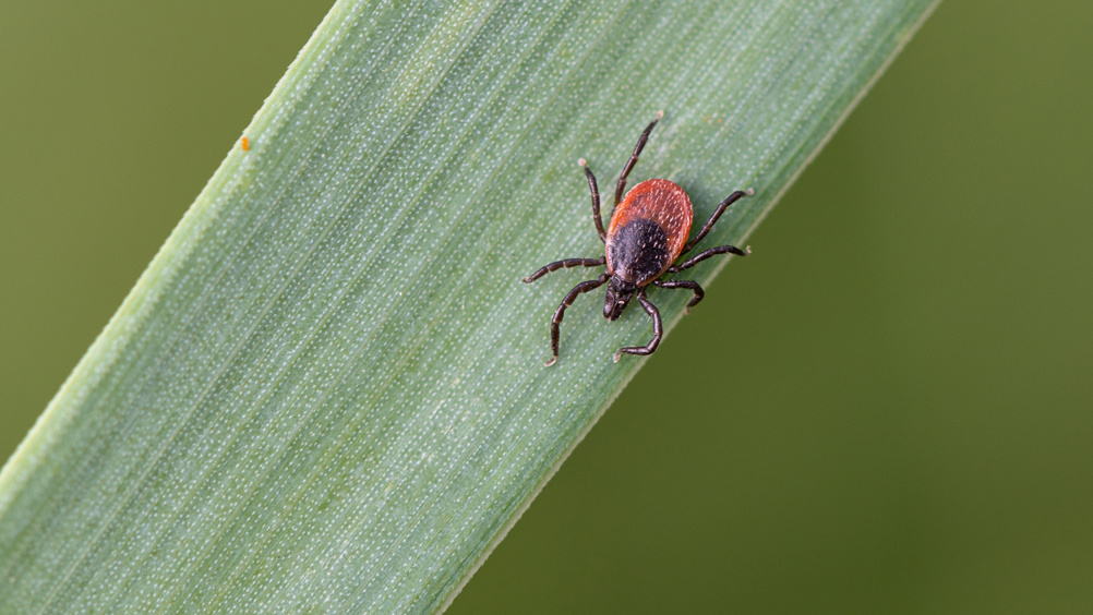
Beyond mammalian hosts, arthropods have been suggested as potential vectors for C. burnetii transmission. The Nine Mile strain of C. burnetii was first isolated in 1935 from the tick Dermacentor andersoni in Montana (Davis et al, 1938), and since then C. burnettii or at least its DNA has been identified in over 40 hard bodied and 14 soft bodied species of tick (Eldin et al, 2017). The prevalence of C. burnetii in ticks appears to be geographically variable and as yet there is no evidence of C. burnettii DNA detection in ticks within Northern Europe (Körner et al, 2021).

Human-to-human transmission

Human-to-human transmission of C. burnetii can occur but is rare. As with livestock, birth products from infected women may be a source of infection, to both hospital staff (Raoult and Stein, 1994), and other pregnant women within the same unit (Amit et al, 2014). There is also limited evidence for sexual transmission (Milazzo et al, 2001). C. burnetii is stable in blood samples for at least 6 weeks at 1–6°C (Kersh et al, 2013), though the risks of transmission via blood products has been estimated to be low (Oei et al, 2014).

Coxiellosis and Q fever within the UK

Coxiellosis

C. burnetii is endemic in the UK. There is evidence for its presence in livestock, wildlife and companion animals. Dairy cattle in particular demonstrate a particularly high herd prevalence with up to 80% of British (Velasova et al, 2017) and over 48% of Northern Irish (McCaughey et al, 2010) dairy farms showing serological evidence of exposure detected in bulk milk samples. The prevalence amongst UK sheep and goats is estimated to be much lower at 10% and 3% of sheep and goat holdings respectively (Lambton et al, 2016). Despite this relatively high seroprevalence, reported numbers of diagnosed cases of infection and of abortion attributed to C. burnetii remain low. However, accurate diagnostics of ruminant abortion, particularly bovine, remains challenging and a diagnosis is not reached in the majority of submissions (Mee, 2020). A retrospective study of livestock placenta samples from abortion events submitted to regional Animal and Plant Health Agency labs in 2010 revealed the presence of C. burnetii DNA in 7 out of 124 cattle and 1 of 9 goat abortion samples but none of the 194 sheep samples tested (Pritchard et al, 2011). These findings of C. burnetii DNA in placenta samples do not confirm attribution of coxiellosis as the cause of the abortion. However, the findings highlight the importance of appropriate biosecurity precautions around the handling of ruminant abortion material and birth products. Another potential reason for the apparent lack of symptomatic coxiellosis in the UK may be due to genetic differences in C. burnetii types found in different livestock species (and also different geographic regions). During the Netherlands outbreak it was determined that goat- and sheep-associated types were the cause of both human clinical Q fever and cases of cattle abortion (Tilburg et al, 2012a; 2012b). Conversely the dominant strains present in cattle did not appear to be associated with overt cattle disease or human infection (Tilburg et al, 2012a; 2012b). Although the C. burnetii types responsible for the disease outbreaks in the Netherlands have been identified in British livestock abortion samples (Hemsley et al, 2023), demonstrating that they are present.

Q fever

The number of diagnosed cases of Q fever across the UK is relatively low, with most cases related to exposure on farms, or to livestock or livestock products (Halsby et al, 2017). While most human Q fever cases in the UK are sporadic in nature, occasional outbreaks have been reported periodically since the 1950s (Harvey et al, 1951). Most are directly associated with livestock exposures such as in Scotland in 2006 where 138 abattoir workers were affected (Pollock et al, 2007). Where direct contact with livestock is not a recognised factor in human case investigations, aerosolised transmission has been suggested such as the 1989 Birmingham outbreak where 147 diagnosed cases of acute Q fever were reported within a 44-mile square area of the city, which was traced back to a farming intensive area where outdoor lambing was taking place, after particularly strong winds (Hawker et al, 1998). On a more local level, disturbance of contaminated straw board during renovations was hypothesised by health officials to be the most likely cause of an outbreak in a factory complex in South Wales in 2002 (van Woerden et al, 2004).

Despite the infrequent nature of outbreaks, there is evidence that at least in some parts of the UK there may be high levels of human exposure. A cross-sectional study of 265 working age patients within a rural general practice in Wales, identified a seroprevalence of 7.9% overall, with farmers exhibiting a significantly higher seroprevalence (15.1%) than non-farmers (4.2%) (Davies et al, 1997). Similarly in a large population-based survey of 2850 individuals carried out in Northern Ireland, a mean seroprevalence of 12.8% was observed (McCaughey et al, 2008) with farmers again exhibiting a significantly higher seroprevalence than the overall population, with 49% demonstrating antibodies to the infection (McCaughey et al, 2008). Highlighted within this study was the observation that seropositive women were significantly more likely to have experienced a still-birth or miscarriage (McCaughey et al, 2008). Despite these findings, it has to be borne in mind that other risk factors, including exposure to other zoonotic infections, cannot be discounted, and the data on Q fever as a cause of human pregnancy loss remain ambiguous. Further studies investigating targeted cohorts of pregnant women in the UK have not observed the same correlations of C. burnetii exposure and adverse pregnancy outcome (Baud et al, 2009; Wheelhouse et al, 2022). Despite the absence of definitive evidence for a role of Q fever in pregnancy complications, there remains a potential risk and appropriate care should be taken in reducing the exposure of pregnant women to livestock (Plummer et al, 2018).

Treatment and prevention of Q fever and coxiellosis

Treatment and prevention strategies in livestock are aimed at reducing rates of abortion and bacterial shedding. The primary method for prevention of clinical disease is vaccination. Commercial vaccines are thought to be most effective when administered to naïve non-pregnant animals, and there is less evidence that vaccination has significant effects on reducing abortion or shedding of previously infected animals (Guatteo et al, 2008; De Cremoux et al, 2012). There has been some limited work on the effects of antibiotics. While oxytetracycline administration has been suggested to have some positive impact on reducing abortion in animals when administered in the latter stages of pregnancy to sheep within a flock with a history of Q fever (Astobiza et al, 2010), there is little evidence that antibiotic treatment reduces bacterial shedding in either cattle or sheep (Astobiza et al, 2010; Taurel et al, 2014).

C. burnetii can be shed in vaginal secretions and birth fluids as well as sporadically in faeces (Guatteo et al, 2007; Roest et al, 2012). Efficient composting is thought to be sufficient to inactivate the organism in faeces prior to application on land (van den Brom et al, 2015b). In lambing/calving pens, good hygiene practice is essential and over 90% of reduction in infectivity can be achieved with prolonged (over 30 minutes) exposure to disinfectants based on hypochlorite, quaternary ammonium or hydrogen peroxide (Plummer et al, 2018). For humans, the primary source of risk is around handling of livestock birth products, even of those animals that are apparently uninfected. Appropriate personal and protective equipment including gloves, protective clothing and respiratory protection should be used when lambing or calving, with frequent attention being paid to hand washing after handling animals (Plummer et al, 2018; Winter et al, 2021).

Conclusions

C. burnetii the causative agent of coxiellosis and Q fever is endemic in livestock across the UK. The number of reported human and livestock cases of disease remain low; however, many cases are asymptomatic and the often-mild symptoms of the disease, described frequently as ‘flu-like’, may be mistaken for other infections. It is therefore likely that the number of animal and human clinical cases are higher than currently reported and there is a real need to investigate exposure and clinical impacts in human populations, particularly those handling livestock and livestock products. Vaccination of livestock may offer a long-term control strategy, but while there is strong evidence that it is effective in the control of clinical disease within flocks/herds, it does not remove the risk of shedding and exposure. Therefore, while we currently do not have a full assessment of risk, as with other zoonoses, it is imperative – particularly at times of lambing/calving – that suitable precautions are taken including the use of personal protective equipment.

KEY POINTS

  • Coxiella burnetii (the causative agent of Q fever) is endemic in UK livestock.
  • In April 2021, livestock Coxiellosis became a reportable disease.
  • The number of reported cases of human Q fever and livestock coxiellosis in the UK is low.
  • Handling livestock or livestock products are the single largest risk factors for Q fever infection in humans.
  • Control of animal infection is primarily via vaccination of livestock.