

Johne's disease is an infectious enteric disease caused by the bacterium Mycobacterium avium subspecies paratuberculosis (MAP). The disease is insidious with a long incubation period which results in chronic inflammation, leading to a progressive granulomatous enteritis. Subsequent poor absorption of nutrients and reduced metabolic efficiency lead to reduced reproductive performance, progressive weight loss, impaired immunocompetence and ultimately reduced productivity.
The disease deleteriously impacts animal welfare and is responsible for economic losses because of decreased productivity, increased replacement costs associated with premature culling and increased mortality. Paratuberculosis also arouses concerns regarding public health; ongoing contention exists around the potential zoonotic link between Johne's disease and Crohn's disease in humans.
Molecular diversity of Mycobacterium avium subspecies paratuberculosis
The two main groups of MAP strains are ‘type S’ and ‘type C’, named after the host species from which they were originally isolated, sheep and cattle respectively (Collins et al, 1990). This nomenclature used for the S- and C-type strain classification implies an exclusive correlation between strain type and host species of origin, which is not the case. C-type strains are capable of infecting a broad range of host species, including sheep, and a wide range of wild and domestic species, including non-ruminants globally (Stevenson, 2015; Bryant et al, 2016). C-type usually predominate in cattle isolates (Stevenson, 2015), although Verdugo et al (2014) reported that S-type strains were more common in New Zealand beef cattle than C-type indicating that S-type also has the capacity to infect cattle and other species as well as sheep (Whittington et al, 2001; de Juan et al, 2006; Ghosh et al, 2012; Verdugo et al, 2014).
In Australia, ovine Johne's disease (OJD) is almost exclusively caused by the S strain (Moloney and Whittington, 2008). In contrast, a study involving seven European countries found that C-type strains were frequently isolated from sheep (Stevenson et al, 2009). Potential explanations for this could be differences in management practices, relative scale of proportions of different livestock species co- or sequentially grazing and evolutionary origins of MAP bacteria populations from livestock imports into Australia and New Zealand (Stevenson et al, 2009). Further, apparent differences in prevalence may be, at least in part, a result of the relatively greater difficulty of culturing S-type isolates, which may have led to underestimations of this strain and microbiological bias in some studies.
The geographical distribution of different MAP strains has likely been influenced over time by numerous factors, including strain virulence, animal movements and farming management practices. At present, whole genome sequencing has shown little supporting evidence for geographically distinct strains although this may change as more MAP isolates are sequenced. One relevant exception is the pigmented type I strains (belonging to the S strain sub-lineage), which seem to be largely restricted to certain geographic pockets within the UK (Stevenson, 2015) and are associated with yellow discolouration of the intestinal mucosal lining. More work is warranted to further our understanding of the genetic diversity of MAP strains within UK livestock populations.
Clinical signs and economic impacts of disease
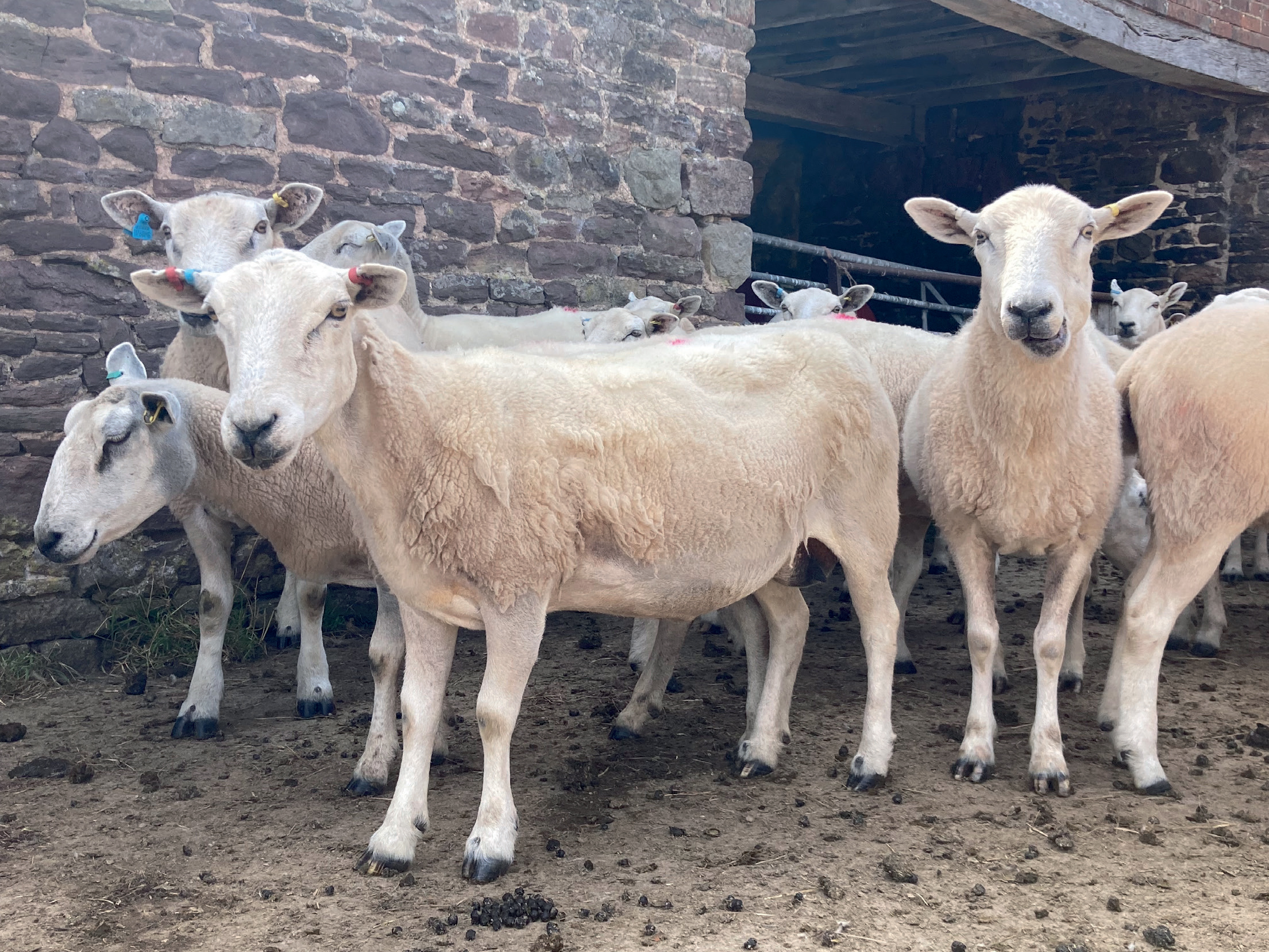
The predominant clinical sign of OJD is progressive weight loss resulting in poor body condition, which is ultimately fatal if affected animals are not culled (Figure 1). Other clinical signs that may be observed include submandibular or ventral oedema as a result of protein losing enteropathy and poor fleece quality (Grieg, 2000). Notably, profuse watery diarrhoea is not a pathognomonic hallmark of Johne's disease as it is in cattle and only a relatively small proportion of sheep with clinical disease will have soft faeces (Grieg, 2000; Windsor, 2015). Clinical disease tends to manifest in adults despite the majority of affected animals becoming infected during the first few months of life (Kreeger, 1991). Onset is slow and gradual with non-specific clinical signs often occurring late in the disease process. However, sheep as young as a year old have been diagnosed with OJD and so it should be considered as a potential differential diagnosis in ill-thrifty shearlings as well as older adults (Scotland's Rural College (SRUC), 2020). Interestingly, some animals appear to be inherently resistant to MAP infection and do not succumb to disease despite being exposed to infective doses of the bacteria (de Silva et al, 2018).
A recent study funded by the Agriculture and Horticulture Development Board (AHDB) found that the presence of MAP in pooled faecal samples correlates with a significant reduction in the longevity of the ewe flock and a significant increase in the replacement costs (Worsley and Davies, publication in prep). Negative effects on fertility have been reported in dairy sheep (Kostoulas et al, 2006) where older animals of parity five and above generated an average of 19% fewer live lambs born per ewe compared with matched uninfected controls. The negative impact of OJD on reproductive performance is likely to be a result of the ewes experiencing a negative energy balance in the pre-clinical phase of the disease as malabsorption leads to progressive malnutrition. When experiencing malnutrition reproductive performance is down-regulated to conserve energy for more essential physiological functions. This manifests as failure to become pregnant and these non-pregnant ewes are culled as unproductive animals without a positive diagnosis being sought; the poor productivity being attributed to a wide range of other causes. This is supported by recent study results (Worsley and Davies, in prep). This highly insidious presentation would explain the enormous discrepancy between the detectable prevalence of MAP at the flock level and the low awareness of the disease among farmers. The premature culling of ewes for poor fertility results in a higher replacement rate than would otherwise be required. The higher replacement rate and reduced longevity results in a younger age profile of the flock and as young ewes are less fecund than mature ewes, this younger age profile inevitably results in reduced flock productivity.
Other plausible associated costs of disease include reduced cull ewe value of thin animals. A project by Meat and Livestock Australia (MLA, 2005) showed that OJD-infected Merino sheep may begin to lose weight nearly a year prior to death from the disease. Infected animals lost an average of 4% of their bodyweight (approximately 1.5 kg) by 8 months before death and by the time of death affected sheep were 32% lighter (approximately 12 kg) than their OJD-free counterparts. The same study also estimated the average OJD-related mortality rate on infected flocks to be 6.2%, ranging widely between 2.1 and 17.5% on different properties. Although increased on-farm mortalities and associated fallen stock costs will no doubt contribute to OJD-associated losses on infected UK farms, it is very likely that mortality rates attributable to OJD are significantly less than those seen in Australia, which may be largely as a result of the reported increased susceptibility of fine-wool Merino type breeds to the disease (Begg et al, 2017).
Transmission
Transmission is predominantly faecal-oral in nature, with young animals being the most susceptible. Infection may occur via ingestion of MAP present in faecal material shed from infected animals, particularly while suckling teats of an infected dam, and also via exposure to MAP-contaminated pasture, forage, feed supplements or water (Windsor and Whittington, 2010). Infection may also occur as a result of ingestion of contaminated colostrum or milk as MAP is also excreted in the colostrum and milk of infected sheep (Lambeth et al, 2004; Nebbia et al, 2006). Shedding of MAP in faeces increases as the disease progresses but can be intermittent and inconsistent especially in the early sub-clinical stages. However, MAP will typically be detectable in faeces prior to antibodies being detectable in blood or milk samples.
In utero infection has also been described and similarly to cattle, is more likely to occur during the clinical versus sub-clinical stages of the disease (Lambeth et al, 2004). MAP has also been isolated from semen although venereal transmission is considered to be of relatively minimal importance compared with other transmission routes described (Eppleston and Whittington, 2001).
Cross-species transmission
There is growing evidence that cross-species transmission of MAP can occur (Stevenson et al, 2009, Verdugo et al, 2014). Considering that MAP is able to survive on pasture for up to 55 weeks (Whittington et al, 2004), this risk is particularly relevant to the UK, where co- or sequential grazing of cattle and sheep is commonly practiced, as is winter (‘tack’) grazing of sheep on dairy farms that have been treated with slurry and farm yard manure. There is also evidence of the same MAP strains being present in wildlife and domestic livestock species on the same farm, although the exact role of wildlife in the maintenance of MAP infections in domestic stock remains unclear (Stevenson et al, 2009). In addition to simply demonstrating interspecies transmission can occur, the frequency, ease and relative risk of transmission, both between domestic livestock and wildlife species, remains to be determined.
Early evidence of interspecies transmission of S-type strains was demonstrated when infected sheep were exported from Germany to Iceland, where S-type strain infection passed to the local cattle population and subsequently back to the sheep population after a depopulation and restocking programme (Fridriksdottir et al, 2000; Whittington et al, 2001). More recently a study on New Zealand farms by Verdugo et al (2014) reported evidence for the occurrence of interspecies transmission if different species are kept in close contact. This study found the same S subtypes in beef cattle and sheep co-grazing on the same farms, and also reported evidence for contact-dependent transmission between deer and beef cattle. Thus, it would seem that co-grazing two or more ruminant species increases the risk of interspecies transmission, providing these species are susceptible to the circulating MAP strain in question.
Conversely in Australia, C-type and S-type strains of MAP have been reported to cause epidemiologically distinct infections (Moloney and Whittington, 2008). The risk of natural transmission of S-type strains from sheep to cattle was thought to be low and only likely to occur when susceptible animals were exposed to high infective doses of MAP (Moloney and Whittington, 2008).
The role of wildlife
While MAP has been isolated from a wide range of UK wildlife species, only lagomorphs and wild ruminants such as deer show evidence of disease as determined by the presence of gross or microscopic lesions with associated acid fast bacteria (Beard et al, 2001a, b). These species have the capacity to actively excrete MAP and as such could constitute wildlife reservoirs (Stevenson et al, 2009). Indeed, a study involving sampling rabbits on beef farms in Scotland concluded that MAP-infected rabbits likely contribute to the persistence of Johne's in domestic livestock and undermine disease control strategies and efforts (Shaughnessy et al, 2013). Identical genotypes, using a combination of techniques, have been obtained from MAP strains isolated from wildlife and domestic ruminants co-inhabiting the same property, which adds weight to the role for wildlife reservoirs in paratuberculosis infection (Stevenson et al, 2009).
The variation in transmission patterns between different countries highlights the need for further research to elucidate inter-species transmission dynamics specific to UK farming conditions, considering local management practices, environmental conditions, numbers and type of livestock and wildlife present, MAP strain populations and interaction with other diseases, which may influence the course of MAP infections.
Diagnosis
Similar to other mycobacterial diseases, diagnostic tests for paratuberculosis have inherently low sensitivity, which is related to the long incubation period when infected animals show no discernible clinical or serological signs and are difficult to detect. However, many diagnostic tests can be useful, practical and cost-effective if they are applied appropriately for a specific purpose or scenario. Limitations of each test and their interpretation need to be understood and communicated effectively between veterinarian and farmer.
Commercially available diagnostic tests for OJD include direct microbiological tests for the MAP pathogen (polymerase chain reaction (PCR) or culture of faeces, milk or tissues or direct visualisation of acid fast bacilli in Ziehl-Neelson stained smears), indirect tests for the host's immune response (antibody detection in milk or serum) or tests assessing the tissue inflammatory response (histopathology and gross pathology). Bacterial culture and molecular techniques such as PCR applied to faecal samples are direct measures of MAP shedding in an individual or pooled group of animals. Serological tests, such as enzyme-linked immunosorbent assay (ELISA), measure the humoral immune response of an individual animal. ELISA can also be used on milk samples, both from individual animals and also on bulk tank samples. These antibody tests are most applicable in the more advanced stages of disease associated with increasingly discernible clinical signs (Nielsen et al, 2008). Despite the low sensitivity in subclinical stages ELISA assays are frequently employed as herd and flock screening tests because of their low cost and practicality. This trade off between cost and sensitivity may be appropriate in certain situations but the onus is on the veterinary surgeon to fully understand and communicate the ramifications of this trade off to the client, especially the high probability of under-diagnosis because of the poor sensitivity of the ELISA. Detection of MAP by PCR assay should be the most sensitive method for flock screening and the addition of a culture step prior to PCR could further increase this sensitivity, but only for those MAP strains that are amenable to laboratory culture techniques. There are currently no independent performance comparisons of the different MAP PCR protocols currently marketed in the UK.
UK prevalence data
Currently, very few estimates of prevalence within the national flock in Great Britain exist. It is very likely that both under-diagnosis, because of lack of awareness, non-specific clinical signs and insufficiently sensitive diagnostic tests, together with underreporting, result in significant under-estimations of flock and within-flock prevalence of OJD in the national flock. The prevalence data sets available are typically from either small samples of flocks (<100), and/or are regionally restricted or unrepresentative because of selection bias.
At the time of writing, the Veterinary Investigation Diagnosis Analysis (VIDA) database reports that 925 positive OJD diagnoses have been made by contributing laboratories over the last decade, the most out of any of the iceberg diseases. OJD diagnoses have been made in all regions of the UK and in almost all counties. VIDA surveillance data are inherently skewed by geographical coverage and submission selection as was the Fallen Stock Project albeit on a larger sample of ewes.
The Animal and Plant Health Agency (APHA) thin ewe investigation project identified OJD in 24 out of 75 English and Welsh farms (32%) based on post-mortem examinations of up to three thin ewes per flock. OJD was by far the most commonly diagnosed iceberg disease in this project (Bell et al, 2021).
The aforementioned AHDB study involving approximately 54 sheep flocks from England, Wales and Scotland, found 64% of holdings had evidence of OJD using pooled PCR (Robinson et al, 2019). Interestingly, none of the farms that were diagnosed with Johne's had previously diagnosed or suspected the presence of the disease in their flocks.
Similar prevalence results were found in a subsequent study (2020) involving 41 Welsh farms. Pooled faecal PCR from 20 ewes on each farm indicated that 29 of the properties had evidence of OJD, putting prevalence estimates at 71% (Worsley et al, in prep). A larger scale project in 2021 involving 59 flocks across England, Wales and Scotland also estimated that approximately 60% of flocks showed evidence of MAP infection using pooled faecal PCR (Worsley et al, in prep).
Control and prevention
The aim for OJD-free flocks is to maintain their status via biosecurity measures that prevent the introduction of the disease onto their farm. For flocks with OJD, the aim is to reduce the number of new infections, as well as maintain biosecurity to prevent reinfection from other infected flocks.
The three main approaches that are used alone or in combination to manage the impacts of OJD are:
- Implement husbandry strategies to reduce the transmission of MAP
- Test and cull programmes to identify and remove positive animals that act as a source of infection
- Vaccinate replacement animals to increase the resistance to infection over time and reduce shedding and thus infectious pressure (Windsor, 2015).
Management strategies
An Australian randomised longitudinal field trial involving 840 Merino sheep naturally exposed to MAP on pasture found that both age at first exposure and the level of exposure to MAP were major determinants of lesion development. This supports a primary focus on limiting exposure of young animals to MAP and reducing the levels of contamination with the bacterium on the pasture (McGregor et al, 2012). In keeping with this aim, prompt culling of clinical cases (either confirmed OJD positive or suspect animals with deteriorating body condition score) is recommended to reduce the residual environmental reservoir of infection since sheep with clinical OJD shed huge numbers of MAP in their faeces, estimated at 108 organisms per gram of faeces (Whittington et al, 2000).
Furthermore, given the very high likelihood of vertical transmission from clinically affected sheep to their progeny, it would be strongly advisable to cull any reproductively active female animals displaying clinical signs of OJD (Lambeth et al, 2004).
Biosecurity measures also need to be in place to prevent further entry of MAP on to a property. This involves keeping a closed flock as much as feasibly possible and ideally sourcing rams and any necessary replacements from flocks that are either OJD free or are on an established vaccination programme. In the UK, only a very small number of flocks are currently either vaccinating or routinely testing for OJD, so sourcing low risk replacements with any level of assurance is likely to be challenging. One breeding company has adopted vaccination of all breeding rams pre-sale but this approach is uncommon at present.
Other potential OJD management interventions, akin to those widely used for Johne's control in both dairy goat and cattle herds, could include ‘snatch’ lambing (whereby lambs are immediately removed from their dam at or as soon as possible after parturition), feeding these lambs on MAP-free colostrum and milk (pasteurised or artificial milk replacer) and rearing replacements in MAP-free locations isolated from the main infected flock, all the while practicing scrupulous farm hygiene and maintaining strict biosecurity between the infected and naïve groups (Djønne, 2010; Windsor and Whittington, 2010; Windsor, 2015). These measures, if they are to be implemented effectively, are extremely labour and time intensive, likely to result in less efficient use of land (more space required to effectively manage two separate groups) and are unlikely to yield immediate results. They may be feasible for small flocks where intensive individual management is possible (e.g. small high-value pedigree and/or dairy flocks), but are unlikely to be applicable to the majority of commercial prime lamb producers or extensive hill sheep enterprises in the UK.
Test and cull
In order for a test and cull programme to successfully halt transmission and reduce the number of new cases, Johne's positive animals need to be identified early enough in the course of infection to allow these infected animals to be removed prior to commencement of MAP faecal shedding (Windsor, 2015). Current diagnostic tests available are generally of low sensitivity and multiple tests performed intermittently over a prolonged period of time are necessary to confer confidence in the true Johne's status of an individual animal or flock/herd (Windsor, 2015). While this is possible in dairy herds, where multiple milk samples can be tested during lactation, there is no available, cost-effective alternative for sheep flocks. Even in the dairy industry this repeated testing is used in addition to other transmission control measures. It is important that producers appreciate that singular negative test results do not guarantee freedom from disease. A risk level accreditation scheme is available in the UK through the Premium Sheep & Goat Health Scheme (PSGHS) managed by SRUC. Membership of the Johne's scheme is extremely low currently, far smaller than the better known maedi visna accreditation scheme. However, in contrast to MAP accreditation schemes, such as the SheepMAP program in Australia, the PSGHS relies primarily on the less sensitive ELISA blood test rather than pooled MAP PCR or culture tests. Given the low sensitivity of the ELISA test combined with the smaller size of the typical UK flock compared with their Australian equivalents it is unlikely that the PSGHS will be able to achieve the same high degree of confidence in the actual disease status of the flock.
Vaccination
In contrast to management interventions or test and cull programmes, vaccination has been shown to be a highly effective way to control OJD in Australia (Juste and Perez, 2011). The vaccine GudairTM (Virbac Ltd, UK) provides very effective and economic flock-level control of clinical disease in a range of Australian flock management systems and underpins the country's national OJD control programme along with a risk-based trading system (Windsor, 2006; Bush et al, 2008; Windsor 2014). Preceding widespread use in Australia, the vaccine was used successfully in controlling disease following an OJD outbreak in Iceland (Fridriksdottir et al, 2000).
It is important to note that vaccination does not entirely prevent infection in all animals, although it does significantly reduce and delay the occurrence of clinical cases and also the rate of MAP shedding from infected animals (Windsor, 2006, 2014).
In accordance with the product datasheet, Gudair is recommended for use in young replacement lambs between 4 weeks to 6 months of age. A single dose is administered with no further boosters required. Vaccination of replacements should continue annually for several years beyond the point at which all animals in the flock have been vaccinated, and unvaccinated animals have been culled (Windsor, 2014). This advice is based on the assumption that a proportion of vaccinated sheep will continue to excrete MAP to some degree, thus maintaining a reservoir of infection in the environment (Dhand et al, 2013; Windsor, 2013).
The value of vaccinating adult sheep is somewhat unresolved. While vaccination of adult sheep and goats in infected flocks and herds can reduce the number of animals developing clinical disease (Corpa et al, 2000), Australian field studies report that significant losses of adult vaccinates will continue in heavily infected flocks that may render ‘whole flock’ vaccination uneconomical (Windsor, 2015).
As a result of the tissue provoking adjuvant, swelling around the injection site is a common side effect and is often prolonged and retracted. If necrosis is extensive this may lead to extra carcass trimming being required at the abattoir. Swelling may affect grazing especially if done when lambs are very young (smaller tissue mass to accommodate inevitable swelling).
Gudair is formulated using the 316F strain which is a C-type strain and yet is reported to be efficacious against S-type strains which predominate in Australia. This evidence of cross reactivity supports the theory that use of the vaccine in the UK should provide good cross protection irrelevant of what strains are present in our flocks. Gudair stimulates both cell-mediated and humoral immunity and is thus unsuitable for differentiating infected from vaccinated animals (DIVA), if a serological test for OJD diagnosis is used as part of test-and-cull strategies or animals tested for export.
Conclusion
In recent years stronger evidence has emerged to show that Johne's is both common in the UK sheep industry and an important cause of insidious, costly, production losses. It is underdiagnosed at present and should be included as part of veterinary investigations into ill-thrift, infertility and poor performance in ewes. Validation and standardisation of commercially available screening tests is needed to improve sensitivity and reproducibility of these assays.
Control of the disease is challenging because of transmission between livestock species and wildlife as well as the long latency and asymptomatic shedding of MAP bacteria into the environment where it can survive and persist for prolonged periods. Clear communication and synergistic actions between farmers, veterinary surgeons, diagnostic laboratories, industry stakeholders and governing bodies are needed to improve awareness of the disease and successful control.
KEY POINTS
- Ovine Johne's disease is currently under-investigated but is likely to be very common and widespread in UK sheep flocks, and a significant cause of lost production. It should be routinely considered as a differential diagnosis for low body condition score, wasting and poor fertility.
- There is growing evidence that cross-species transmission between cattle and sheep occurs in other countries and more work is warranted to investigate inter-species transmission dynamics specific to UK farming conditions.
- Serology antibody tests are useful for diagnosis of symptomatic animals but too insensitive to be useful for screening flocks or individuals. Faecal polymerase chain reaction (PCR) is more appropriate but variation between laboratories remains a concern.
- Control of the disease is challenging because of the long incubation period with asymptomatic faecal shedding of Mycobacterium avium subspecies paratuberculosis (MAP) during this time, ability of the bacteria to survive in the environment for prolonged periods and reservoirs of MAP infection in local wildlife populations.
- Vaccination has proved to be a highly effective control measure in other countries and more practical than test and cull or artificial rearing alternatives. It is under-utilised in the UK at present.